online learning with Doc note master|| Himanshu paneru
Streptococcus pyogenes
Streptococcus pyogenes are gram positive cocci.
SYSTEMIC BACTERIOLOGY
8/3/20242 मिनट पढ़ें


STREPTOCOCCUS PYOGENES
Streptococcus pyogenes is a species of Gram-positive bacteria, it is gram positive cocci.
streptococci is typically found in chains or pairs.
It is an important cause of bacterial infections worldwide
Morphology
· Gram-positive bacteria
· Shape: spherical or oval
· Size: About 0.5 to 1.0 µm in diameter.
· Arrangement: Arrange in chain.
· Capsule: Some strains of possess capsule.
· Motility: Non-motile
· Spore: Non-sporing
Pathogenesis
Streptococcus pyogenes produce pyogenic infection that spread locally along with lymphatic & blood stream.
Ø Source of infection – Contaminated food, lesion, infected blood or body fluid such as saliva, wound, or nasal secretions, etc.
Ø Mode of transmission – transmitted by direct or indirect content, ingestion & inhalation.
Ø Route of transmission – respiratory tract, GI tract, etc.
Ø Incubation period – about 3 to days.
A. SUPPRATIVE INFECTION:
1) RESPIRATORY INFECTION –
Sore throat (acute tonsillitis and/or pharyngitis) is the most common streptococcal diseases.
Tonsillitis is more common in older children and adults.
2) Skin & soft tissue infection –
Strep. Pyogenes causes subcutaneous infection of skin lymphangitis & cellulitis.
It include wound & burn.
It may lead to fetal septicemia.
Strep. Pyogenes is also known as ‘FLASH EATING BACTERIA’.
Bacterial infection such as Erysipelas & impetigo.
3) Streptococcal toxic shock syndrome –
TSS is a condition in which the entire organ system is collapse, leading to death.
4) Genital infection –
Aerobic & Anerobic streptococci are normal inhabitant of female genital tract.
Important causative agent of puerperal sepsis.
5) Other supprative infection
Strep. Pyogenes may cause abscess in internal organ such as brain, lungs, liver & kidney.
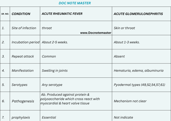
B. NON-SUPPRATIVE INFECTION:
Lab diagnosis
Ø Specimen:
Throat swab
Wound swab
Pus
CSF
Blood
Ø collection & transport:
Specimen should be collected on sterile container.
Pike’s medium is used for transport
Ø Microscopy:
Gram staining - Gram positive bacteria seen in chain formation.
Ø Culture:
Best grow in blood agar at 37®C for 24-48 hours in presence of 5 – 10% CO2.
Ø Colony morphology:
Colony of strep. Pyogenes are-
Shape – round
Size- 0.5-1mm
Surface – smooth
Color – grey
Opacity – translucent
Ø Biochemical reaction:
Camp test – negative
Catalase – negative
PYR test – positive
Ø Serological test:
Anti streptolysin o (ASO) is most widely used
Ø Antigen detection test:
ELISA
Agglutination test
Ø Molecular method:
Polymerase chain reaction
Ø Antibiotic susceptibility testing:
Oral penicillin V or amoxicillin
Prevention
Good hygiene practices such as handwashing and covering the mouth and nose when coughing or sneezing.
Avoiding close contact with infected person.
Proper wound care and management to prevent skin infections.


